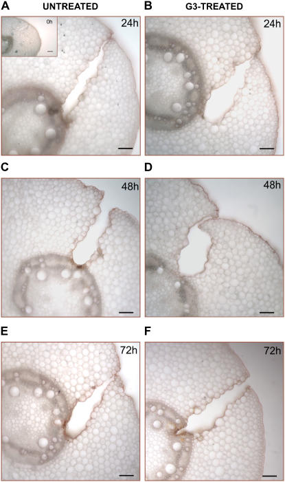
Figure 10.
Light microscopy detection of suberin aliphatic domain in wound-healing maize mesocotyls. Histochemical analysis was performed in hand-cut cross sections (approximately 100 μm thick) obtained from the wounded zone of the mesocotyl of G3-untreated and G3-treated plants, at 0 (inset), 24 (A and B), 48 (C and D), and 72 (E and F) h after wounding. Sections were preincubated for 10 min in 50% ethanol and then stained for 20 min in a filtered saturated solution of Sudan IV in 70% ethanol. After washing in 50% ethanol (1 min), sections were observed under light microscopy. Bars = 100 μm.

