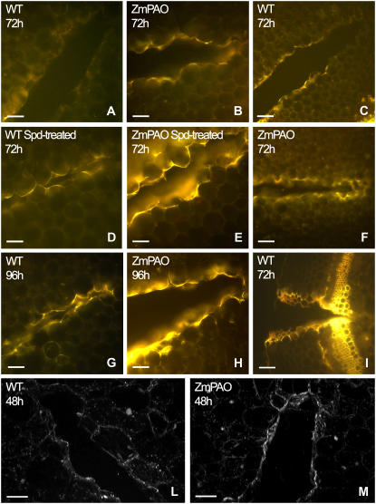
Figure 11.
Blue-induced autofluorescence and laser scanning confocal microscopy analysis in wound-healing tobacco plants overexpressing ZmPAO in the cell wall. Histochemical analysis under fluorescence microscopy was performed in hand-cut cross sections (approximately 100 μm thick) obtained from the wounded zone of the second internode (numbering from the shoot apex) of Spd-untreated (A–C and F–I) and Spd-treated (D–E) wild-type (WT) as well as ZmPAO transgenic tobacco plants overexpressing ZmPAO in the cell wall (ZmPAO), at 72 and 96 h after wounding. Sections were directly mounted on slides and observed for autofluorescence under blue light (A–I). A, B, D, E, and G to I, bar = 100 μm; C and F, bar = 200 μm. Confocal microscopy analysis was performed on hand-cut cross sections from wild-type and ZmPAO transgenic tobacco plants at 48 h after wounding. L and M sections show a three-dimensional reconstruction of autofluorescence images after blue excitation. L and M, bar = 100 μm.

