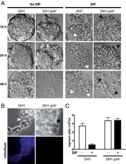
Figure 2.
iplA mutant cells showed almost complete inhibition of DIF-induced autophagic cell death. (A) Vacuolization was inhibited in DH1.iplA−(ins) mutant cells subjected to DIF. Wild-type and DH1.iplA−(ins) mutant cells were starved for 8 h, and then they were washed and further incubated in the presence of 100 nM DIF (DIF; right) or in its absence (No DIF; left). DIC microscopy pictures at 16, 24, or 48 h after the initial 8-h starvation period. Vacuoles (white arrowheads) were visible in DH1 wild-type cells subjected to DIF. However, there were almost no vacuoles in the absence of DIF or in mutant iplA− cells subjected to DIF. The latter group showed paddle cells (Levraud et al., 2003) (black arrowheads) occurring most often at the periphery of cell aggregates. (B) Cellulose shells did not occur in DH1.iplA−(ins) mutant cells in response to DIF. After 16 h of incubation in DIF, DH1 wt, and DH1.iplA−(ins) mutant cells were labeled with calcofluor. For each cell type, pictures taken by phase-contrast (top) or fluorescence (bottom) microscopy corresponded to the same microscopic field. Arrowheads as in A. Only DH1 wild-type, not DH1.iplA−(ins) mutant cells were vacuolized and calcofluor positive. (C) Clonogenic cell death was markedly less in DH1.iplA−(ins) mutant cells than in DH1 wild type cells in response to DIF. After 24 h of incubation with or without DIF, HL5 rich medium was added, and cells were counted after a further 48-h incubation period.

