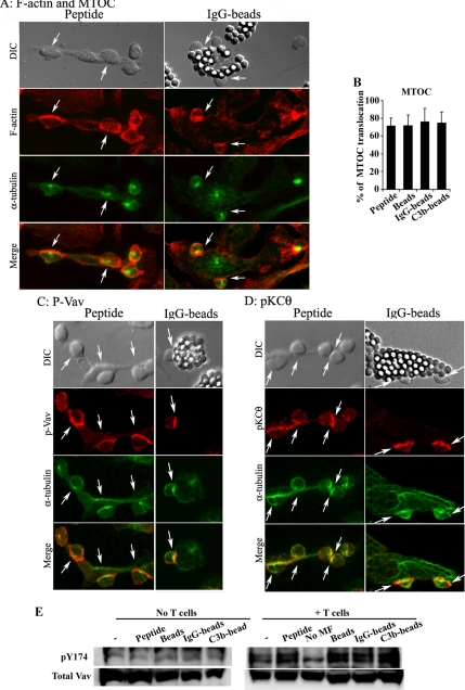
Figure 5.
Phagocytic macrophages induce T-cell F-actin clustering, MTOC translocation, and recruitment of TCR proximal signaling proteins. Conjugates formed with macrophages loaded with OVAp (left panels) or OVA IgG-beads (right panels) are shown. DIC images are shown in the top panels. Cells were stained for F-actin with phalloidin (A); for phosphorylated Vav (C), or PKCθ (D; red), and for the MTOC, using the α-tubulin antibody (green). Merged images are shown in the bottom panels. Arrows indicate T-cell conjugates positive for MTOC and other molecules. (B) The number of conjugates in which the T-cell MTOC was translocated was quantified and expressed as the percentage of the total number of conjugates formed. Results are presented as the arithmetic means ± SD of five experiments; statistical comparisons were made with the Mann-Whitney test (*p < 0.05). (E) Immunoblots showing phosphorylated Y174 Vav and total Vav expression in macrophages loaded with 1 μM of OVA peptide or OVA differently opsonized to beads (left) and in conjugates formed between T-cells and these macrophage populations (right).

