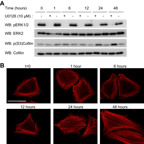
Figure 6.
MEK signaling regulates actin stress fibers and cofilin phosphorylation. WM793 cells were incubated with the MEK inhibitor U0126 (10 μM) or an equal volume of dimethyl sulfoxide (DMSO) (−) for 1, 6, 12, 24, and 48 h. (A) Cells lysates generated at the indicated times were analyzed by Western blotting for levels of phospho-ERK1/2, total ERK2, phospho-(S3)-cofilin and total cofilin. Shown are representative blots from three independent experiments. (B) Cells were fixed at indicated times and stained with TRITC-phalloidin to visualize F-actin. Bars, 50 μm.

