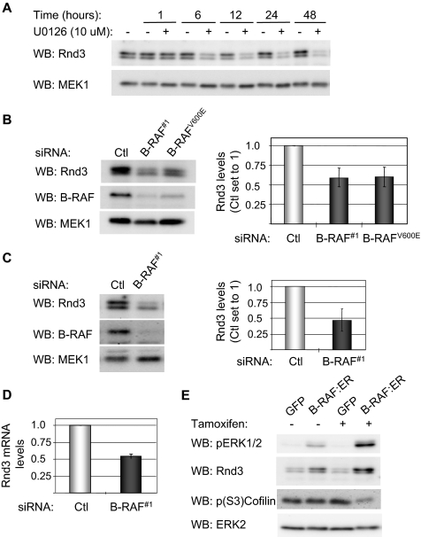
Figure 7.
B-RAF and MEK regulate the expression of Rnd3. (A) WM793 cells were treated with 10 μM U0126 or equal volume DMSO for increasing times, as indicated. Cell lysates were western blotted using antibodies to Rnd3 and total MEK, as a loading control. (B) Western blot analysis of Rnd3, B-RAF, and MEK1 levels in WM793 cells transfected with control, B-RAF#1, or B-RAFV600E siRNA. Data are the mean ± SD for the Rnd3/MEK1 ratios from three independent experiments. The control siRNA condition is set to one. (C) Similar to above, except that WM115 cells were transfected with control or B-RAF#1 siRNA. (D) Total RNA was extracted from control and B-RAF knockdown WM793 cells. Quantitative RT-PCR analysis was performed with primers specific for Rnd3 and actin. The graph represents the mean percentage of change in Rnd3 mRNA relative to actin from two independent experiments. (E) NHEM cells infected with adenovirus to express ΔB-RAF:ER* or GFP were incubated 18 h in the absence or presence of tamoxifen. Cell lysates were Western blotted for phospho-ERK1/2, Rnd3, phospho-(S3)-cofilin, and total ERK2.

