Figure 3.
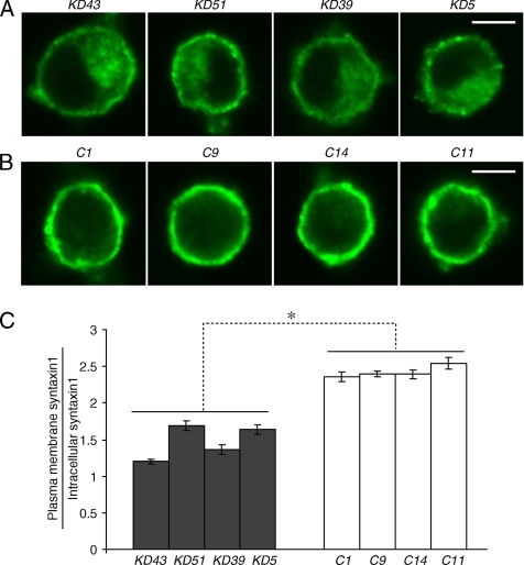
Confocal immunofluorescence microscopy reveals syntaxin mislocalization in Munc18-1 knockdown cells. (A and B) Representative single-cell confocal images of cells from four pairs of knockdown (A) and control (B) that were stained by anti-syntaxin1 antibodies (HPC-1) followed by Alexa 488–conjugated anti-mouse antibodies. Scale bar, 5 μm. (C) The graph shows the proportion of syntaxin1 found in the plasma membrane to that found inside the cell for four pairs of knockdown clones and their paired controls. Error bar, SEM (n = 14–26). *p < 0.01; a statistically significant difference.

