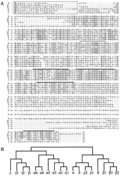
Figure 1.
Human GABA-A receptor θ subunit. (A) Alignment of the deduced amino acid sequences of the human GABA-A receptor α1(18), β1(18), γ1 (19), δ (unpublished results), ɛ (4), and θ subunits. Numbering of amino acids is given by assigning the initiating methionine as 1. Positions where amino acid residues are conserved in four or more sequences are shaded. Putative signal peptides (20) are boxed, putative transmembrane domains are overlined, and the two cysteine residues conserved in the ligand-gated ion channel family are joined by a solid line. (B) Dendrogram of the deduced amino acid sequences of the GABA-A receptor (and ρ1–ρ2 of GABA-C) family. The analysis was performed with clustalw (GCG).

