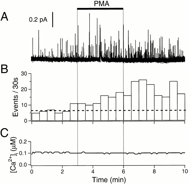
Figure 7.
Stimulation of exocytosis by PMA. (A) Amperometric record. The cell was preincubated in 50 μM BAPTA-AM for 1 h, and then loaded with dopamine and indo 1-AM. PMA (100 nM) was applied for 3 min as indicated by the bar. (B) The rate of exocytosis for the same cell. The horizontal broken line indicates the average rate of exocytosis in the control period. (C) Simultaneous Ca2+ measurement using indo-1 dye in the same cell.

