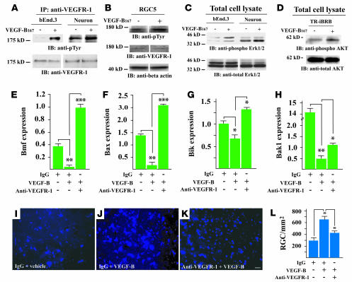
Figure 6. VEGFR-1 mediates the effect of VEGF-B.
(A) VEGF-B167 stimulation resulted in VEGFR-1 activation in the bEnd.3 cells and the cortex neurons using the immunoprecipitation assay and anti–VEGFR-1 antibody, followed by the Western blot assay using anti-phosphotyrosine antibody (anti-pTyr). VEGFR-1 was detected in bEnd.3 cells and cortex neurons using Western blot assay. (B) VEGF-B167 induced VEGFR-1 activation in RGC5 cells using Western blot assay and antibodies against phosphotyrosine and VEGFR-1. (C) VEGF-B167 treatment led to ERK1/2 activation in both bEnd.3 cells and cortex neurons using Western blot assay and antibodies against phosphorylated and total ERK1/2. (D) VEGF-B treatment induced Akt phosphorylation in TR-iBRB cells using Western blot assay and antibodies against phosphorylated and total Akt. (E–H) VEGFR-1 neutralizing antibody treatment abolished to various degrees the inhibitory effect of VEGF-B on the expression of Bmf (E), Bax (F), Bik (G), and Bak1 (H) in RGC5 cells. (I, J, and L) VEGF-B protein treatment increased RGC survival in the ONC-injured retina. Scale bar: 20 μm. (I, K, and L) VEGFR-1 neutralizing antibody treatment largely abolished the VEGF-B–induced RGC survival in the ONC-injured retina. *P < 0.05, **P < 0.01, ***P < 0.001.

