Figure 3.
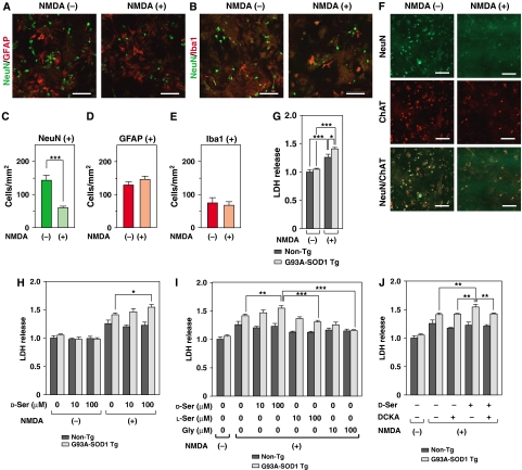
Elevated vulnerability of PSCs from ALS mice to toxicities of D-Ser/NMDA. (A–F) NMDA treatment on PSCs triggers neuronal death, but not glial death. Double IF analysis with mouse monoclonal anti-NeuN (green) antibody in association with rabbit anti-GFAP (red (A)) or rabbit anti-Iba1 (red (B)) antibody was performed at 24 h after the beginning of treatment with 500 μM NMDA on PSCs from non-Tg mice. (C–E) The numbers of NeuN- (C), GFAP- (D), and Iba1- (E)-positive cells were counted in five random visual fields and averaged (***P<0.001; error bars represent s.e.m.). (F) NeuN-positive cells were characterized by double IF analyses with anti-ChAT antibody at 24 h after treatment with NMDA. Most NeuN-positive cells were ChAT-positive. Few PSCs were NeuN-, GFAP-, and Iba-1-negative. Scale bars=100 μm (A, B, F). (G–J) Cell mortality of PSCs from non-Tg and ALS mice was determined with LDH assays. The extent of LDH release was shown as folds of values measured in cells from non-Tg without NMDA treatment. (G) Treatment with 500 μM NMDA caused a 1.41-fold increase of LDH release from PSCs of ALS mice, whereas a 1.26-fold elevation was observed in those from non-Tg (N=3). (H) Treatment with 0, 10, and 100 μM of D-Ser under 500 μM NMDA on PSCs from ALS mice exhibited 1.41, 1.46, and 1.55-fold increase, respectively (N=3). (I) The effects of L-Ser or Gly on 500 μM NMDA-induced toxicity were measured and compared with that of D-Ser treatment (N=3). (J) The effect of treatment with an antagonist to the glycine sites of the NMDARs, DCKA (30 μM), on D-Ser (100 μM)/NMDA (500 μM)-induced toxicity was examined (N=3). (*P<0.05, **P<0.01, ***P<0.001; error bars represent s.e.m.). Shown is a typical result of an experiment repeated three times.

