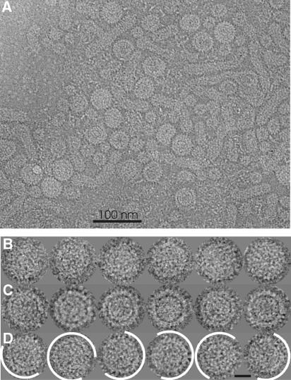
Figure 1.
Micrograph and gallery of HBV embedded in vitrified buffer. (A) Part of a micrograph (inverted contrast) recorded with a Philips CM120 Biotwin. (B) Gallery of selected particles with compact morphology. (C) Gallery of selected particles with gapped morphology. (D) Gallery of particles with mixed morphology. Areas consistent with the gapped morphology are delineated in white. Particles appear bright against darker background. The length of the bar in panel D equals 20 nm.

