
Figure 1. Spontaneous voltage changes recorded from IC granule cells under conditions of current clamp.
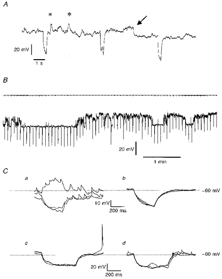
A, an extract of a membrane potential recording from an IC neurone in a slice, held hyperpolarized by a steady current of 10 pA of injected current. The periodic hyperpolarizations (every 5 s) are in response to additional superimposed 0.5 s current injections of 5 pA. On the trace are marked two putative synaptic events (*) and a ‘step’ change in membrane potential from -61 to -70 mV (arrow); see text for further details. B, from a different cell in a semi-isolated clump of neurones, a longer trace of membrane potential record with accompanying current record showing the level of steady clamping current (-7.5 pA) and at 0.2 Hz the imposition of 0.5 s, 5 pA hyperpolarizing current injections. The membrane potential wavered between preferred values: the more negative level was -79 mV. C, responses captured in two further neurones in response to 0.5 s current injections of 10 pA (Ca and b) or 15 pA (Cc and d). In Ca the record was noisy and small spikelets appeared on the recordings, whereas in Cb which was recorded 2 min after Ca, the voltage trace was relatively quiet and the spikelets were no longer observable; each panel shows 3 consecutive superimposed traces. Cc and d, 3 superimposed voltage traces are shown in each panel, showing responses to hyperpolarizing current in a cell which displayed both full-sized action potentials (Cc) and spikelets (Cd) which appeared 3 min later. Steady holding currents for the two cells, which were both in clumps of IC neurones, were - 5 pA (Ca and b) and -18 pA (Cc and d).