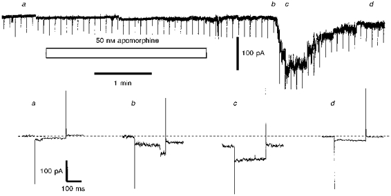
Figure 4. Apomorphine induces a delayed inward current characterized by a stepwise increase and decrease.

The upper part of the figure shows a chart record of the current required to hold an IC neurone at -80 mV; every 5 s the cell was stepped to -100 mV to assess whole-cell conductance and the traces so generated at the timesindicated (a-d) are displayed in the lower half of the figure. Apomorphine (50 nm) was applied as indicated by the bar. Note that prior to apomorphine exposure the neurone displayed small current fluctuations (cf. Fig. 2) and a clear transition is resolved in trace b: the magnitude of the latter conductance change was 606 pS. The ACSF contained 0.5 μm TTX and the preparation was a slice preparation.
