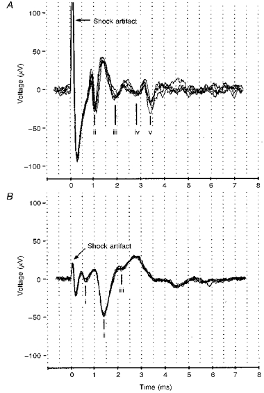
Figure 1. Compound action potentials recorded in retina evoked by optic tract stimulation.

A, four negative waves labelled ii, iii, iv and v are typically recorded at latencies of 1, 1.8, 2.8, and 3.4 ms. B, a different recording of compound action potentials showing the fastest wave, i, at a latency of 0.7 ms, in addition to waves ii and iii.
