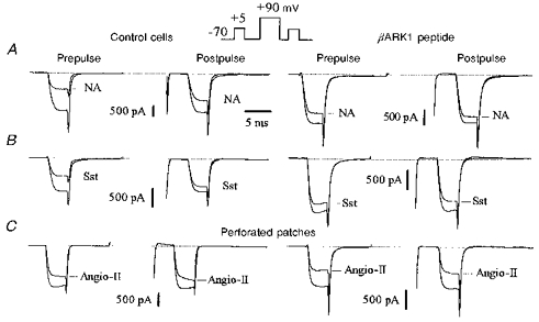
Figure 1. The C-terminus of βARK1 reduces ICa inhibition by noradrenaline and somatostatin but not by angiotensin II.

Superimposed Ca2+ current traces recorded in the absence or presence of noradrenaline (NA; 10 μm, A), somatostatin (Sst; 500 nm, B) and angiotensin II (Angio-II; 500 nm, C) in uninjected neurones (left panels, Control cells) and in neurones preinjected with 200 μg ml−1 of the βARK1 construct (right panels). Ca2+ currents were elicited by the double-pulse protocol as illustrated in the inset. The outward currents elicited by the conditioning voltage pulse to +90 mV (at break) are omitted. In this and subsequent figures we have referred to the test depolarizations before and after the conditioning voltage pulse as ‘prepulse’ and ‘postpulse’, respectively. Responses to angiotensin II in C were recorded using the perforated-patch method where access resistances were 10.5 MΩ (left traces) and 8 MΩ (right traces). ICa inhibition was measured at steady state, 4 s after application of noradrenaline or somatostatin and 15–18 s after application of angiotensin II. Cells were recorded 48 h after injection. In this and subsequent figures, the horizontal dotted lines at the top of the traces indicate the zero current level.
