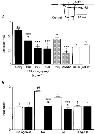
Figure 3. βARK1 peptide expression prevents voltage-dependent inhibition of ICa.

A, bar graph summarizing the effects of βARK1 peptide expression on ICa inhibition induced by noradrenaline (10 μm, filled bars), somatostatin (500 nm, shaded bars) and angiotensin II (500 nm, open bars). Uninj, uninjected control cells. Bars represent means ±s.e.m. for the number of cells indicated. Somatostatin and angiotensin II inhibition was recorded in cells preinjected with 200 μg ml−1βARK1 construct. Cells were recorded 48 h after injection. *** P < 0.0001, compared with the respective controls. Inset, ICa amplitude was measured isochronally 4 ms after the onset of the test pulse (dashed line) from the zero current level obtained in the presence of cadmium (Cd2+, 500 μm). ICa was elicited from −70 to +5 mV. B, bar graph summarizing the effects of the βARK1 peptide on the facilitation ratio (postpulse : prepulse) in the absence or presence of neurotransmitters as indicated. □, control; ▪, βARK1 peptide. *** P < 0.0001.
