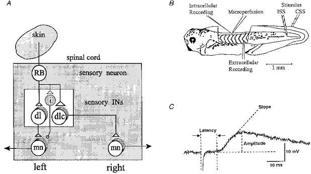
Figure 1. The sensory pathways and methods used.

A, simplified diagram of the pathways from a skin sensory Rohon-Beard (RB) neuron on the left side to motoneurons (mn) on both sides of the spinal cord of the Xenopus tadpole. The pathway from the sensory RB neuron to the contralateral motoneuron is via the dorsolateral commissural (dlc) interneuron (IN) and to the ipsilateral motoneuron is probably via dorsolateral (dl) interneuron. Open triangles represent glutamatergic excitatory synapses. The small shaded neuron (i) is the proposed new class of inhibitory interneuron that makes glycinergic synapses (small shaded circle) with ipsilateral motoneurons (see Discussion). B, diagram of the experimental preparation and arrangement of electrodes to stimulate both sides of the tail skin and record responses of a spinal motoneuron. For details see Methods. C, measurements made on the stimulus-evoked EPSPs. The onset latency is the time from the stimulus artefact to the start of the EPSP. The amplitude of the EPSP was measured from the baseline (resting potential) to its peak. Slope is a straight line fitting the rising phase of the EPSP reflecting the rate of rise (in mV ms−1).
