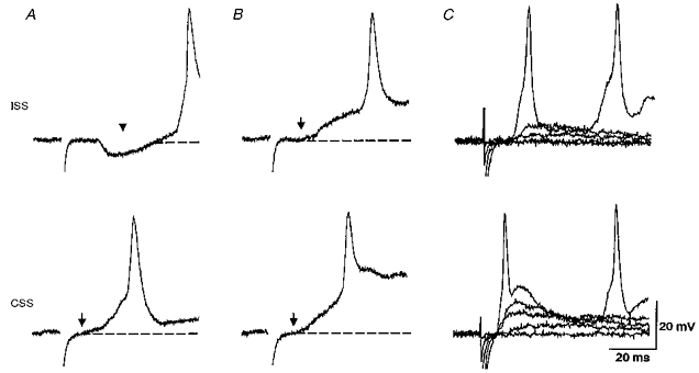
Figure 2. Responses of motoneurons to skin stimulation on the ipsi- (ISS) and contra (CSS)-lateral sides of the tail and the effects of strychnine.

A, the response to ISS (at artefact) shows an obvious IPSP (at arrowhead) before EPSP and action potential. The response to CSS shows an EPSP (onset at arrow) which rises quickly to an action potential as swimming starts. Dashed lines represent resting membrane potentials. B, during microperfusion of 2 μM strychnine the ISS-evoked IPSP is blocked. This reveals an underlying EPSP (at arrow) and the latency of the action potential becomes shorter. The EPSP and action potential in response to CSS shows no change and the spike latency is still shorter than the ISS-evoked spike. C, in strychnine, grading the skin stimulus from below threshold reveals differences in both the EPSP and spike latencies evoked by ISS and CSS. A and B were from a neuron at the 6th segment (resting potential, −70 mV); C was from another neuron at the 7th segment (resting potential, −70 mV).
