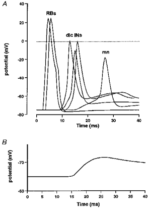
Figure 9. Simulated membrane potential responses of selected neurons in the pathway from the skin to a contralateral motoneuron.

A, after the RBs are stimulated (2 are shown firing), there is a progression in the excitation of dlc interneurons depending on their distance from the site of stimulation (3 are shown). The EPSPs that these produce sum in the motoneuron and in this case reach threshold to initiate a spike in the motoneuron. B, an example of a simulated motoneuron EPSP (cf. Fig. 2B) as used for measurements.
