
Figure 11. Light microscopic morphology of recorded neurons as revealed by biocytin labelling and three-dimensional reconstruction by a confocal laser scanning microscope.
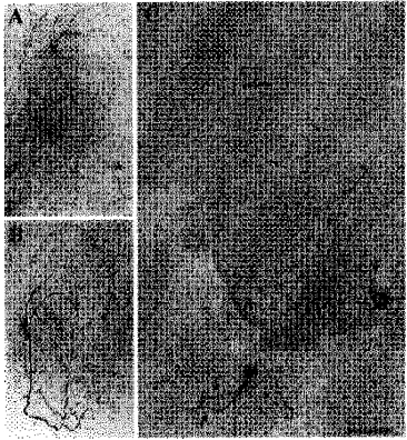
A, cluster I neuron having an axon projecting to the area of the paraventricular nucleus of the hypothalamus (marked by arrow) and a few local axon collaterals in the SCN (marked by asterisks). B, cluster III neuron with a characteristic large axon collateral network within the SCN. The collaterals carried large numbers of varicosities and coursed through the ventral SCN. No projection axon was identified in this cell. C, cluster II neuron having an axon projecting to a site dorsal to the SCN (thin arrow). This cell presents a clear example of varicose dendrites (thick, short arrows) and also possesses a small number of axon collaterals (marked by asterisks). Scale bar: A, 130 μm; B, 65 μm; C, 75 μm.