
Figure 12. Morphology of cluster I neuron with somatic spines and axon collaterals originating from distal dendritic segments.
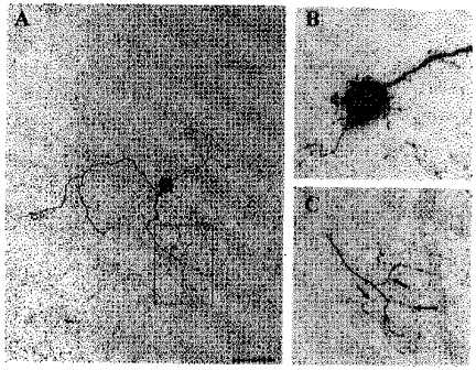
This dendritic configuration is referred to as ‘dendroaxon’. The cell was reconstructed as in Fig. 11. A, overview of the neuron with soma, dendrites and axon collaterals. B, partial reconstruction of the soma and proximal dendrites showing numerous spines or prolonged appendages on both soma and dendrites. C, enlargement of the box shown in A, revealing axon collaterals (marked by arrows) originating from a distal dendrite. Note the fine calibre of the axons. Scale bar: A, 40 μm; B, 14 μm; C, 20 μm.