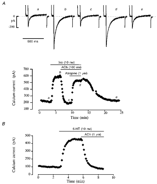
Figure 2. Accentuated antagonism of ACh on ICa in human atrial myocytes.

The cells were first superfused with control solution and then exposed to the drugs during the periods indicated in the graphs by the bars. A, superfusion of the cell with 10 nM Iso produced a nearly maximal stimulation of ICa, which was fully abolished by 100 nM ACh. Addition of the muscarinic antagonist atropine (1 μM) restored ICa to an amplitude which was close to the Iso-stimulated level. Upon washout of the drugs and return to control conditions, ICa slowly recovered its control amplitude. The individual current traces shown in the upper part of the figure were obtained at the times indicated by the corresponding letters in the graph. The dotted line indicates the zero current level. B, application of 10 nM 5-HT to another myocyte also produced a strong stimulation of ICa. Application of 1 μM ACh in the continuous presence of 5-HT induced a complete inhibition of the serotoninergic response.
