
Figure 6. Enhancement induced by ATPγS dialysis occludes 17β-oestradiol potentiation of kainate-induced currents.
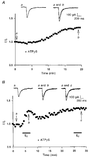
A, intracellular dialysis of ATPγS (500 μM) increased the amplitude of kainate-induced currents. The illustrated example shows that the increase occurred ≈10 min after ATPγS dialysis and gradually reached steady state at ≈20 min. B, the illustrated whole-cell recording was obtained from a CA1 neurone responsive to ATPγS (500 μM) dialysis. Application of 17β-oestradiol (100 nM, 3 min) during the initial period of recording elicited a rapid potentiation in the peak currents. The potentiation was partially reversible upon removal of 17β-oestradiol. After the amplitude of the currents increased by ATPγS dialysis reached a steady level, addition of 17β-oestradiol caused no further potentiation. In A and B, actual current traces corresponding to the specific time points (arrows) are displayed at the top.