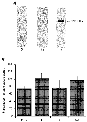
Figure 2. Absence of involvement of iNOS in the L-NMMA enhancement of ICa.

A, Western blot experiment showing the absence of iNOS protein on isolated ventricular cells. Cells were lysed soon after, or 24 h after, dissociation, as described in Methods. The probe used was a monoclonal antibody anti-murine macrophage inducible NOS. 0 = lysate at time 0, 24 = lysate at 24 h and C = positive control (lysate of murine macrophages previously treated with interferon-γ and LPS for 12 h). B, bar graph showing no substantial variation in the L-NMMA (1 mM) effect in cells used both soon after (1, within 5-6 h), and the day after dissociation (2). The bar labelled 1 + 2 is the effect on all cells tested, and Dexa represents the cells isolated and kept in the presence of 3 μM dexamethasone in the Tyrode solution from the start of the procedure and throughout the storage period.
