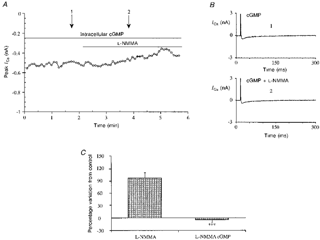
Figure 6. Lack of effect of L-NMMA in the presence of intracellular perfusion with cGMP.

A, time course of ICa from a typical experiment with L-NMMA (1 mM) in the presence of cGMP. Stimulation protocol as in the previous figures. The patch electrode contained the standard intracellular solution described in the Methods section with 10 μM cGMP. B, single traces of ICa at the times indicated by the arrows in A: control (1) and L-NMMA (1 mM) (2). C, bar graph showing the lack of effect of L-NMMA in the presence of cGMP (10 μM, ***P < 0.0005 vs. no cGMP) compared with the effect in its absence.
