
Figure 2. Spatially resolved changes in [Ca2+]i in individual CA1 pyramidal neurones during a spontaneous Ca2+ burst.
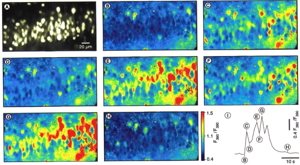
A, an epifluorescence image (taken at 390 nm excitation light) of the cell body layer in the CA1 region of a hippocampal slice from a 3-day-old rat taken with a × 40 objective lens. B-H, pseudocolour ratio images were taken at various time points, as indicated in I, during a Ca2+ burst consisting of four Ca2+ transients. I, time course of the Ca2+ burst calculated as a mean of all individual changes in [Ca2+]i (n= 107 cells).