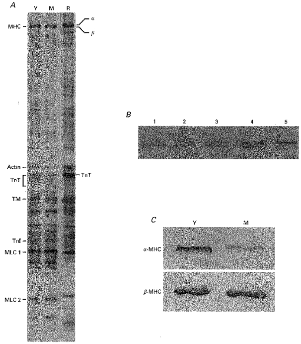
Figure 2. Protein analysis of guinea-pig and rat samples.

A, silver-stained SDS-polyacrylamide gel from young (Y) and mature (M) guinea-pig hearts and from euthyroid rat heart (R) (0.8 μg protein per lane). Contractile proteins were identified by Western immunoblotting with specific antibodies, as indicated in the text: MHC, myosin heavy chain; TnT, troponin T; TM, tropomyosin; TnI, troponin I; MLC, myosin light chain. B, separation of the two MHC isoforms in rat heart samples. Lane 1, hypothyroid; lane 5, euthyroid; lanes 2–4, 3:1, 1:1 and 1:3 mixtures of hypo- and euthyroid rat samples, respectively. C, although in guinea-pig ventricular muscle the α- and β-MHCs were not separated by SDS-PAGE (A, lanes Y and M), the presence of α-MHC and β-MHC in young and mature guinea-pig hearts could be visualized with Western immunoblotting using two monoclonal antibodies directed specifically against α-MHC and β-MHC (0.8 μg protein was loaded in each lane).
