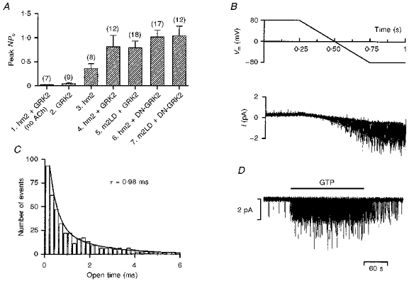
Figure 1. Properties of the muscarinic K+ channel in CHO cells.

A, mean ( ± s.e.m.) NPo from 7 to 18 cell-attached patches on various groups of cells. The peak value of NPo when the pipette was first attached onto a cell was measured. 1, cells transfected with the wild-type receptor and wild-type receptor kinase. ACh was absent from the pipette. 2, cells transfected with the wild-type receptor kinase, but not the receptor (ACh present). 3, cells transfected with the wild-type receptor, but not the receptor kinase (ACh present). 4, cells transfected with the wild-type receptor and wild-type receptor kinase (ACh present). 5, cells transfected with mutant receptor and wild-type receptor kinase (ACh present). 6, cells transfected with the wild-type receptor and mutant receptor kinase (ACh present). 7, cells transfected with mutant receptor and mutant receptor kinase (ACh present). All groups of cells were also transfected with the muscarinic K+ channel (Kir3.1/Kir3.4). B, 10 superimposed traces of single channel currents (bottom) during repeated voltage ramps (top; Vm, membrane potential). At positive potentials no channel activity was recorded, whereas there was intense channel activity at negative potentials. Inside-out configuration (at least 2 active channels in patch). C, open time histogram. The number of channel openings of a particular open duration is plotted against the open time; 0.2 ms bin width. The data are fitted with an exponential function with a time constant, τ, of 0.98 ms. The data were collected over a 1 s period about 3 min after the attachment of the pipette. Inside-out configuration (at least 2 active channels in patch). D, single channel currents recorded in an inside-out patch (at least 2 active channels in patch). ACh was present in the extracellular solution in the pipette. When 0.1 mm GTP was applied to the intracellular face of the patch during the period indicated by the horizontal bar, the muscarinic K+ channel was activated and channel activity was observed. Transfection: A1 and A4, stable transfection with DNA for hm2 and GRK2 and transient transfection with DNA for Kir3.1, Kir3.4 and GFP; A2, transient transfection with DNA for GRK2, Kir3.1, Kir3.4 and GFP; A3, stable transfection with DNA for hm2 and transient transfection with DNA for Kir3.1, Kir3.4 and GFP;A5, transient transfection with DNA for m2LD, GRK2, Kir3.1, Kir3.4 and GFP or stable transfection with DNA for m2LD and transient transfection with DNA for GRK2, Kir3.1, Kir3.4 and GFP; A6, stable transfection with DNA for hm2 and transient transfection with DNA for DN-GRK2, Kir3.1, Kir3.4 and GFP; A7, stable transfection with DNA for m2LD and transient transfection with DNA for DN-GRK2, Kir3.1, Kir3.4 and GFP; B-D, stable transfection with DNA for hm2 and GRK2 and transient transfection with DNA for Kir3.1, Kir3.4 and GFP (see Methods for details).
