
Figure 2. Immunodetection of NHE2 and NHE3 in pig and human colonic LMV.
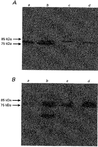
Colonic LMV and homogenate (25 μg of protein each) were separated by 8% SDS-PAGE and electrotransferred to a nitrocellulose membrane. Immunoblotting was carried out as described in Methods. A, immunodetection of NHE2; B, immunodetection of NHE3. Lane a, pig colonic homogenate; lane b, pig colonic LMV; lane c, human colonic homogenate; lane d, human colonic LMV.