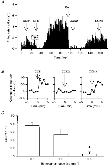
Figure 4. Effects of acute α1-adrenoceptor antagonism on CCK-induced excitation of oxytocin neurones.

A, the firing rate of an oxytocin cell (averaged in consecutive 10 s intervals) in a morphine-dependent rat under urethane anaesthesia administered CCK (20 μg kg−1, i.v.), and infused i.c.v. with benoxathian (5.3 μg min−1, hatched box, Ben), naloxone (NLX; 5 mg kg−1, i.v.) and benoxathian (25 μg, i.c.v., Ben); the benoxathian infusion delayed the peak withdrawal response (see Fig. 3C) and the later injection of benoxathian reversibly inhibited the withdrawal excitation and the response to CCK. B, the change of firing rate of the oxytocin cell in A following each CCK injection (at t = 0 min) on an expanded time scale; the later injection of benoxathian blocked the response to i.v. CCK (CCK2). C, CCK2:CCK1 ratios from morphine-withdrawn and naïve rats treated with 0.3 (n = 3), 1.6 (n = 11) and 5.3 μg min−1 (n = 6) benoxathian i.c.v.; *P < 0.05 compared with the groups administered the lower two doses, one-way RM ANOVA followed by Student-Newman-Keuls analyses.
