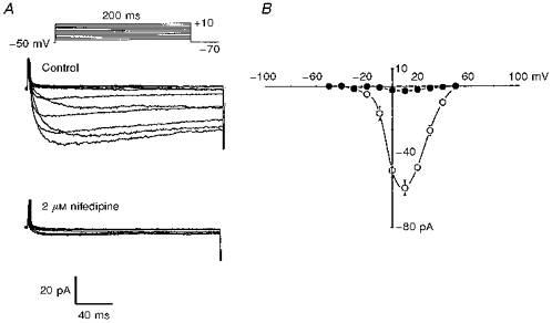
Figure 4. Characterization of macroscopic Ca2+ (Ba2+) currents (ICa) in cat cerebral VSMCs.

A, representative macroscopic Ca2+ currents carried by Ba2+ recorded from cat cerebral VSMCs evoked by 200 ms depolarizing pulses elicited from a holding potential of −70 mV to test potentials between −50 and +50 mV in 10 mV increments, before (upper panel) and after (lower panel) application of nifedipine to the bath. B, average peak current-voltage relation before (^) and after (•) application of nifedipine (n = 6, P < 0.001). Vertical bars denote s.e.m.
