
Figure 5. Apical membrane localization of hPepT1 in human intestine and Caco-2 cells.
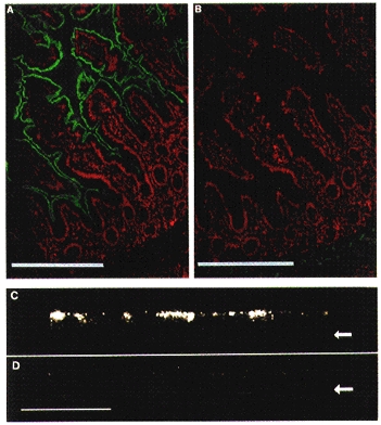
Immunolocalization of hPepT1 by anti-P085 (anti-hPepT1) immunostaining at the apical membrane of human ileum and a Caco-2 monolayer. A and B, immunofluorescence in a section of human ileum. The FITC signal (anti-hPepT1) is shown in green and propidium iodide fluorescence (tissue morphology) is shown in red. The section shown in B was stained in the presence of 5 μg ml−1 peptide P085. Scale bar, 100 μm. A similar staining pattern was observed in human duodenum. C and D, immunofluorescence, imaged by confocal laser scanning microscopy, through Caco-2 cells perpendicular to the plane of the monolayer. D shows a control stained with pre-immune rabbit serum. Arrows indicate the position of the base of the monolayer. Scale bar, 100 μm. Human tissues were stained with affinity-purified anti-P085 antibody at a concentration of 0.05 mg ml−1, as detailed in Methods. Caco-2 cells were stained with anti-P085 polyclonal serum at a 1:40 dilution.