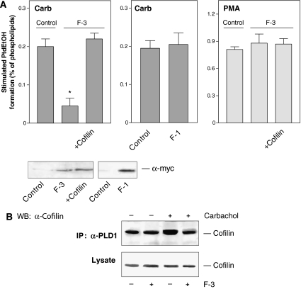
Figure 8.
The F-3 fragment of PLD1 specifically reduces PLD stimulation by carbachol and interaction of PLD1 with cofilin. (A) HEK-293 cells were transfected with empty vector (Control), F-1 or F-3, either alone or together with wild-type cofilin. After 48 h, stimulated [3H]PtdEtOH accumulation was determined in the presence of 1 mM carbachol (Carb) or 100 nM PMA. Data shown are means±s.e. (n=3–4). The immunoblots show expression of c-myc-tagged F-3 and F-1 fragments in cell lysates. (B) HEK-293 cells were transfected with wild-type PLD1 alone or together with the F-3 fragment. After 48 h, the cells were treated for 15 s without (−) or with (+) 1 mM carbachol, followed by cell lysis and immunoprecipitation with the anti-PLD1 antibody. The PLD1 immunoprecipitates (IP) and total lysates were resolved by SDS–PAGE and probed with anti-cofilin antibodies (α-cofilin) as indicated. The results shown are representative of 3–4 experiments. WB, Western blot.

