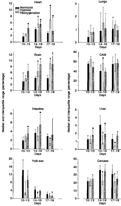
Figure 2. Cardiac output distribution in the chick embryo in normoxia, hypoxia and after 5 min of reoxygenation for three different incubation time groups.

Bars represent median percentage of CO directed to each organ and interquartile range (p25-p75). * Significant difference compared with normoxia (P <0.05); † significant difference compared with normoxia (P <0.01).
