
Figure 7. Current-voltage (I-V) relationships for the NPPB- and glibenclamide-sensitive conductances in the apical membrane.
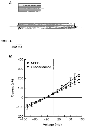
A, a representative tracing of the glibenclamide-sensitive current obtained from amphotericin B-permeabilized monolayers in response to a voltage step protocol from -90 to +120 mV (15 mV step increments) at a holding potential of 0 mV. Glibenclamide (200 μm) was added to the apical solution. B, NPPB- and glibenclamide-sensitive components of the apical membrane current were plotted as a function of voltage. Experiments were performed under conditions where the basolateral surface was permeabilized with amphotericin B and bathed in KMeSO4 Ringer solution with 10 mm NaCl. Standard Ringer solution was used to bathe the apical surface of the epithelium. NPPB or glibenclamide, at a concentration of 200 μm, was added to the apical solution. Mean reversal potentials for NPPB- and glibenclamide-sensitive currents were -28 ± 3 and -27 ± 1 mV, respectively (n= 7, N= 4 for each experiment).