
Figure 5. Single-channel properties measured from three types of transfected cells.
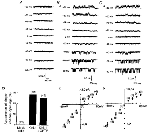
Single-channel current traces were recorded in the cell-attached patches with 140 mm K+ pipette solution, for cells superfused with a 140 mm solution. A, mock-transfected cell. B, Kir6.1-transfected cell. C, cell co-transfected with Kir6.1 and CFTR. Short horizontal bars to the right of each trace indicate the zero current level and numbers to the left of each trace the test potential. Bb and Cb show single-channel I-V relationships. Means ±s.d. Numbers in parentheses are the number of observations. D, percentage appearance of single-channel openings was estimated in each set of cells (mock-, Kir6.1- and Kir6.1-CFTR-transfected) and is summarized in the bar-graphs. Numbers in parentheses indicate the number of trials.