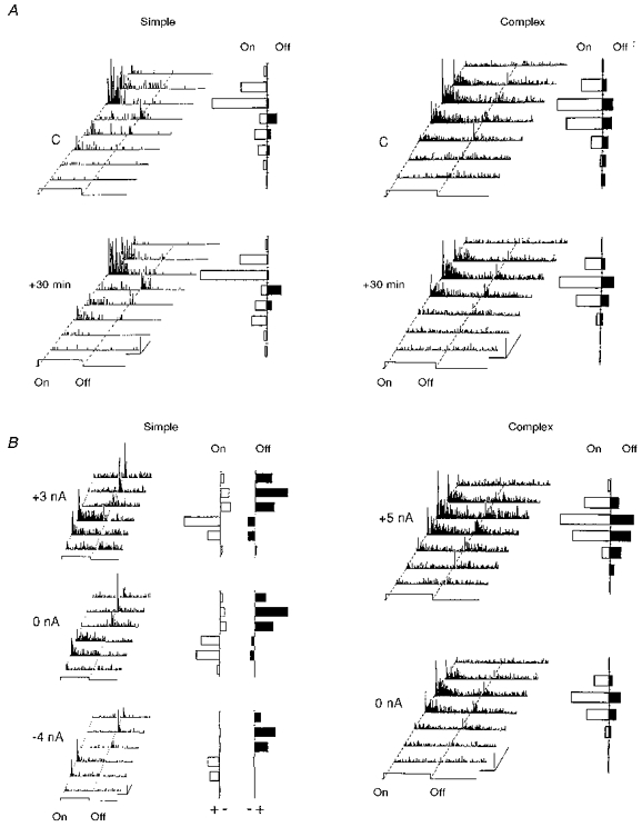
Figure 2. Intrinsic variability of the spatial RF organization.

A, the temporal stability of the spatial profile of ‘on’ and ‘off’ responses was assessed in simple (left panel) and complex (right panel) cells by repetitively exploring the whole extent of the RF with a light bar flashed ‘on’ and ‘off’ in 8 positions, 30 min apart. The cumulative ‘on’ and ‘off’ responses shown for each position (to the right of the corresponding PSTH) remained unchanged. Calibration bars: horizontal, 1 s; vertical, 10 AP s−1; oblique, 1 deg (RF width axis). B, left panel: in order to study the degree of invariance of the spatial RF organization as a function of the level of excitability artificially imposed by a constant iontophoretic current, three RF maps of the same simple cell recorded in an 8-week-old kitten were constructed during the constant application of either positive current (+3 nA), no current (0 nA), or negative current (-4 nA) through the recording pipette. Cumulative excitatory (+) and inhibitory (-) responses were quantified for the ‘on’ (open bars) or ‘off’ periods (filled bars) by subtracting the spontaneous activity (measured for the same temporal window duration between trials) from the visual activity. An increase in early inhibition triggered by the characteristic antagonistic to that of the field under test was apparent when the neuron was held at the more depolarized level. In spite of the large changes in mean level of activity imposed by iontophoresis, the general spatial organization of the RF and the degree of overlap between the ‘on’ and ‘off’ zones remained unchanged whatever the value and polarity of the applied current. Right panel: a complex RF map recorded in a 24-week-old kitten is shown in the control situation (0 nA), and when an iontophoretic current is applied (+5 nA). No inhibitory responses were revealed even under sustained depolarization (+5 nA), in contrast to that observed for simple cells. Calibration bars: horizontal, 1 s; vertical, 10 AP s−1; oblique, 1 deg.
