
Figure 7. Intracellular application of TEA or ShB peptide disrupts endogenous BK channel inactivation.
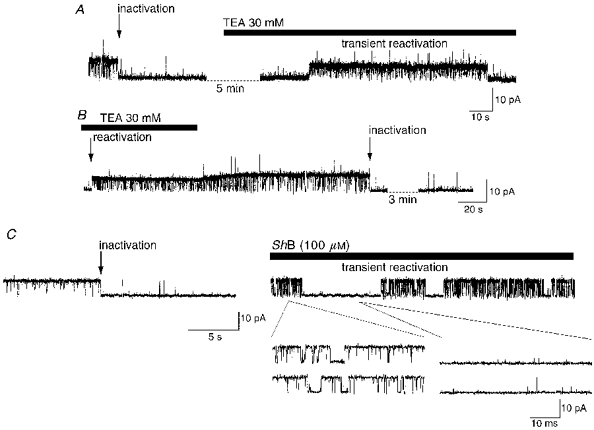
A, after BK channel inactivation at +40 mV (10 μM Ca2+), application of 30 mM TEA to the internal surface of the patch caused a transient reactivation of channel activity. Note the reduced amplitude of events in TEA, both before and after reactivation. B, in patches where a sustained reactivation was produced by intracellular TEA, inactivation (arrow) could be re-established by TEA removal, which can be observed as a progressive increase in single channel amplitude. C, after inactivation of a BK channel at +40 mV, low Po short open time behaviour was observed. Following addition of the ShB peptide (100 μM) to the internal surface of the patch the voltage was stepped to -60 mV for 1 min and then returned to +40 mV. In the presence of the ShB peptide, sustained inactivation was not re-established, instead characteristic short (left-hand expanded traces) and occasional long blocks were observed interspersed with periods of inactivation (right-hand expanded traces).