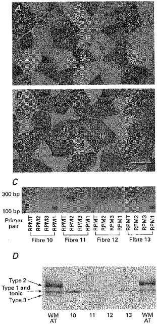
Figure 4. MHC mRNA transcripts and protein isoforms in single fibres obtained from the fast region of the anterior tibialis muscle.

Expression of MHC mRNA transcripts and protein isoforms in immunohistochemically identified frog fibre types obtained from the fast region of the anterior tibialis muscle. A and B, serial sections of anterior tibialis muscle stained for reactivity to the MHC monoclonal antibodies NA8 (A) and F8 (B). Scale bar, 100 μm. C, agarose gels showing expression pattern of the four MHC transcripts in 60 μm segments of immuno-identified fibres (fibre numbers correspond to those shown in A and B), measured by RT-PCR using the four clone-specific PCR primer pairs. D, MHC isoforms measured by SDS-PAGE of 60 μm segments of the same fibres shown in A and B are compared with the banding pattern from whole AT muscle (WM AT). The immuno-classified type 1 fibre (no. 10) expressed only RPM1 mRNA (C) and a single MHC protein band that migrated at the position of the middle band of the triplet seen in whole AT muscle (D). The type 2 fibre (no. 12) expressed only RPM2 mRNA and a single protein band corresponding to the upper band of the triplet. The immuno-classified type 1–2 fibres (nos 11 and 13) coexpressed both RPM1 and RPM2 transcripts and contained 2 MHC protein bands corresponding to the upper and middle bands of the triplet. The upper band (type 2) is barely visible in fibre 11 and was barely detectable in the original gel for fibre 13 and is not apparent in the scanned figure. This banding pattern matches the faint immunoreactivity to NA8 antibody in fibre 13.
