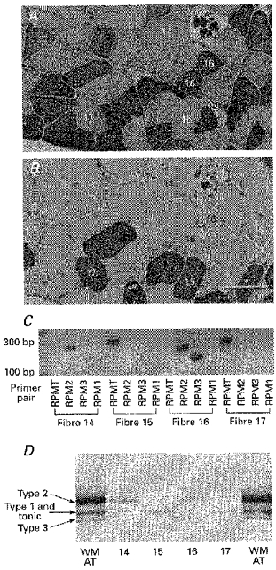
Figure 5. MHC mRNA transcripts and protein isoforms in single fibres obtained from the slow region of the anterior tibialis muscle.

Expression of MHC mRNA transcripts and protein isoforms in immunohistochemically identified frog fibre types obtained from the slow region of the anterior tibialis muscle (labelling scheme as in Fig. 4). A and B, serial sections of anterior tibialis muscle stained for reactivity to the MHC monoclonal antibodies D5 (A) and MHC-S (B). Scale bar, 100 μm. Immuno-classified tonic fibres (no. 15 and no. 17) expressed only the RPMT mRNA (C) and contained a single MHC protein band migrating at the position of the middle band of the triplet seen in whole AT muscle. The type 3 fibre (no. 16) coexpressed RPM3 and RPM2 transcripts and contained 2 protein bands migrating at the position of the upper and lower bands. The type 2 fibre (no. 14) expressed RPM2 and a single protein isoform at the position of the upper band.
