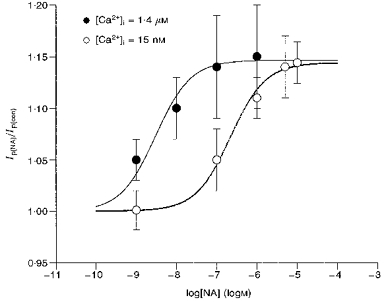
Figure 4. [Ca2+]i affects the sensitivity of the NA-induced stimulation of Ip but not maximal stimulation.

Using the same protocol shown in Fig. 1 but with [Ca2+]i buffered to 1.4 μm, we recorded the stimulation of Ip by 0.001, 0.01, 0.1 and 1.0 μm NA. The mean ratio of Ip(NA)/Ip(con)±s.d. (number of cells) was, respectively, 1.05 ± 0.02 (10), 1.10 ± 0.03 (8), 1.14 ± 0.05 (7) and 1.15 ± 0.03 (7). These data are shown as •. For purposes of comparison we also plot the data from Fig. 1 as ○. Based on the curve fit of a single binding site model (continuous lines) the maximal stimulation of Ip in 1.4 μm[Ca2+]i was 14.7 % whereas in 15 nm[Ca2+]i it was 14.4 %, and the K0.5 in 1.4 μm[Ca2+]i was 3 nm whereas in 15 nm[Ca2+]i the K0.5 was 219 nm. These data show that the maximal stimulation of 14–15 % is independent of [Ca2+]i but the sensitivity to NA increases nearly two orders of magnitude when [Ca2+]i is high. Moreover, the [Ca2+]i independence of maximal stimulation indicates propranolol has effectively blocked β-activation, since at 15 nm[Ca2+]i, β-activation inhibits Ip by about 25 %, whereas at 1.4 μm[Ca2+]i, β-activation has almost no effect at holding voltage of 0 mV. There is no indication of these β-mediated effects in the data shown here.
