
Figure 3. Positive labelling of MNTB neurones by a Kv3.1 antisense probe.
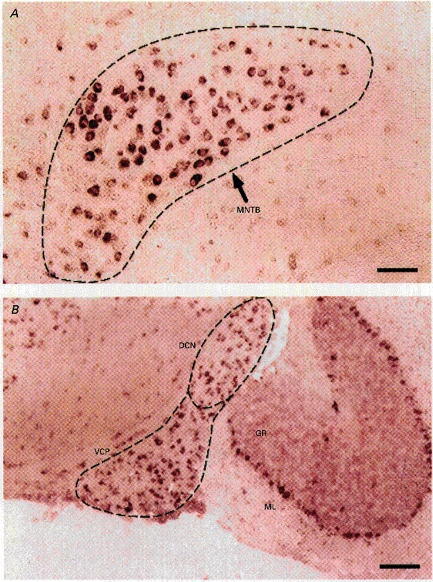
A, strong labelling of the principal neurones of the MNTB region. B, labelling of neurones in the dorsal cochlear nucleus (DCN), ventral posterior cochlear nucleus (VCP), cerebellar granule cells (GR) and Purkinje neurones. The latter are situated between the molecular layer (ML) and the GR layer. This experiment was performed on a 12-day postnatal mouse. Scale bar, 100 μm.