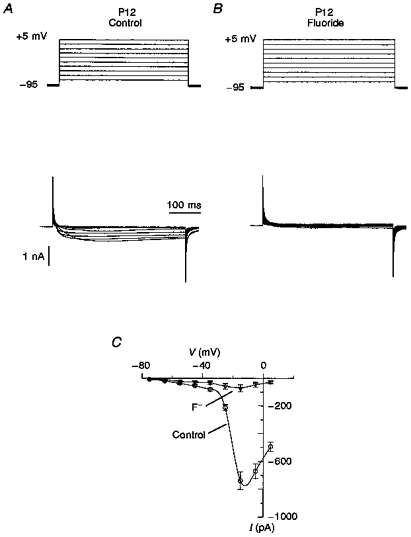
Figure 3. Ca2+ currents in P12 neurones.

A, typical records of Ca2+ current in P12 neurones obtained by membrane depolarization from -95 mV in 10 mV steps. There were no transient Ca2+ currents in the region from -70 to -40 mV. The threshold for activation of Ca2+ currents was about -45 mV. The voltage step protocol is shown at the top. B, Ca2+ currents in the presence of 5 μM F− after a short period (5 min) of intracellular perfusion. Fluoride ions were added to verify whether the LVA Ca2+ current is present in P12 neurones and hidden by the larger HVA Ca2+ current. In general, in the presence of F− ions, 90 ± 5 % of the Ca2+ current was blocked and no LVA current was found. C, I-V relationships of peak currents registered in the absence and presence of F− have only one peak at about -15 mV, corresponding to HVA Ca2+ current.
