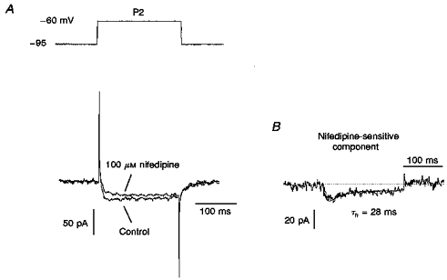
Figure 4. Blocking effect of nifedipine on LVA Ca2+ current in P2 visual cortical neurones.

A, LVA Ca2+ current recorded in P2 neurones before (Control) and after blocking by 100 μM nifedipine. The current was obtained by 35 mV step depolarization from a holding potential of -95 mV (top). B, the nifedipine-sensitive component was obtained by subtracting LVA Ca2+ current after blocking by 100 μM nifedipine from the control LVA Ca2+ current. It was fitted in terms of the m2h model (continuous line) with the inactivation time constant (τh) indicated.
