
Figure 3. The rhythmic IPSPs, but not the tonic hyperpolarization, are mediated by GABAA receptors.
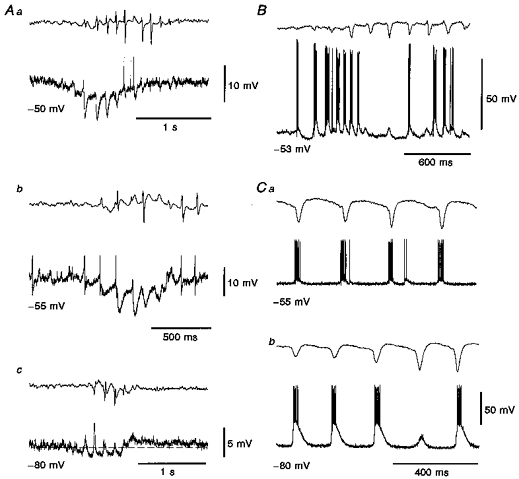
A, intracellular voltage records show the tonic hyperpolarization present during spontaneous SWDs (b and c are from the same neurone). At -80 mV (c) the tonic hyperpolarization and the tonic depolarization present at the end of SWD are clearly visible. B, intracellular activity recorded with a KCl-filled electrode show the lack of any rhythmic hyperpolarizing potentials at -53 mV, and the presence of the tonic hyperpolarization starting well before the first large spike in the EEG. C, intracellular records from another TC neurone recorded with a KCl-filled electrode show full depolarizing envelopes at -55 mV (a), which at -80 mV (b) become very large depolarizations (i.e. EPSP, reversed IPSPs and LTCP) evoking robust action potential bursts. In Aa and b, the amplitude of the action potentials has been truncated for clarity.