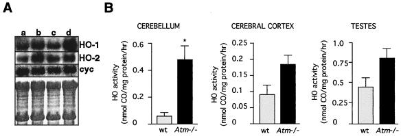
Figure 2.
Increased expression and activity of HO in brains from Atm-deficient mice. (A) Representative Northern blot of 10 μg of total RNA isolated from 3-month-old male littermates as follows: wild-type cerebral cortex (lane a); Atm-deficient cerebral cortex (lane b); wild-type cerebellum (lane c); and Atm-deficient cerebellum (lane d). cyc, Cyclophilin control. The bottom box is the methylene blue-stained blot for assessing RNA loading. (B) Total HO activity in the cerebellum, cerebral cortex, and testes of Atm-deficient (gray bars) and wild-type mice (black bars). Each bar represents the mean ± SEM. Statistical significance of differences between and within groups was assessed with the unpaired t test with the Scheffe correction for multiple comparisons. ∗, Significance with P < 0.05. Three animals of each genotype at 3 months of age were used for the HO activity determinations. The specific activity in wild-type cerebellum was 77 pmol of CO per mg of protein per hr; in Atm-deficient animals it was 458 pmol of CO per mg per hr (P < 0.05). The values for wild-type and Atm-deficient cortex and testes were not statistically significantly different.

