Figure 3.
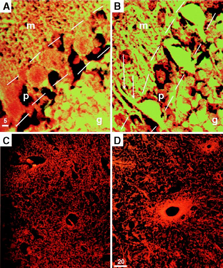
Purkinje cells and cells surrounding blood vessels show increased levels of HO protein in the absence of ATM. The yellow fluorescent signal shown in A and B is from anti-HO-1 staining in a wild-type (A) and Atm-deficient (B) mouse cerebellum at high magnification (×100) to show the significant increase in staining in the Purkinje cell layer. m Indicates the molecular layer, p and outline indicate the Purkinje cell layer, and g indicates the granular cell layer. Note the intense signal in the Purkinje cell soma of the Atm-deficient brain in B. The increased staining in the molecular layer is likely caused by HO in the dendritic arbors, as the spines are well demarcated by the intense staining (double arrows). The granular cell layer (g) staining is similar in the wild-type and Atm-deficient animals. C (wild-type) and D (Atm-deficient) show the increase in bright orange signal from anti-HO-2, which is prominent in cells surrounding the vasculature of the cerebral cortex in the Atm-deficient mouse (D). (×40.)

