
Figure 1. α-BgTx blocked ACh-evoked currents.
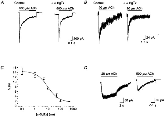
The sensitivity of ACh-evoked currents to α-BgTx was examined by incubating the neurones in the absence (control) or presence of 500 nM α-BgTx for ≥ 15 min before ACh application. A, when macroscopic currents were evoked by high concentrations of ACh (here 500 μM), α-BgTx had little effect on current amplitude. B, the net current elicited by lower concentrations of ACh (here 20 μM) was significantly reduced by α-BgTx, primarily due to a decrease in a later, slow component with little change in peak amplitude. Thus the time course of the current was significantly faster. C, plot of the decay time constant of macroscopic currents elicited by ACh (20 μM) as a function of α-BgTx concentration. The concentration-dependent inhibition by α-BgTx was well fitted by a sigmoidal curve indicating an IC50 of 10.05 nM and an apparent Hill coefficient of 0.995. Each point on this curve was derived from the τ values of single exponential fits of three to nine ACh-evoked currents in the presence of the indicated concentration of α-BgTx. Error bars indicate s.e.m.D,α-BgTx-sensitive component currents. The amplitude and time course of the α-BgTx-sensitive component of ACh-evoked current were obtained by pointwise subtraction of averaged currents recorded in the presence and absence of α-BgTx (see text). ACh-evoked currents were averaged by digitization and alignment at the onset of ACh application (control, n = 7; +α-BgTx, n = 6). The subtracted current revealed that the α-BgTx-sensitive component of the currents evoked by 20 μM ACh had a protracted time course with little desensitization over the 10 s period examined.