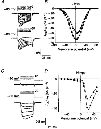
Figure 2. High-voltage-activated VOCCs in differentiated NG108-15 cells.

Recordings were made in Ca2+-Ni2+ containing bath solution (10 mM Ca2+, 0.1 mM Ni2+). Current responses on stepping from the holding potential (Vh = −80 mV) to different test potentials (A and C) are shown with corresponding I-V relationships (B and D) evoked by sequential 5 or 10 mV depolarizing steps from the holding potential (Vh = −80 mV (▪ in B, • in D) and −50 mV (▴)). The two HVA components, L-type (A and B) and N-type (C and D) were pharmacologically separated by 1 μM ωCgTX GVIA (A and B) or 10 μM nifedipine (C and D), respectively.
